Multicolor Flow Cytometers: Immunological & Cell Analysis Tips
Flow cytometry is an innovative and essential technology used in the field of immunology for the analysis of cellular characteristics. This technique revolves around the use of flow cytometers, which enable scientists to analyze the physical and chemical properties of cells or particles that pass through them. Multicolor flow cytometers are a specialized variant that allows researchers to simultaneously measure multiple parameters on individual cells within complex populations.
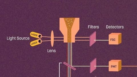
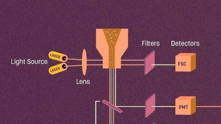
The operation of a multicolor flow cytometer involves several key steps, starting with sample preparation. In most cases, cells of interest are first isolated from a biological sample, which may be blood, tissue, or another fluid. The cells are then treated with fluorescently labeled antibodies that bind specifically to proteins of interest on the cell surface. These antibodies typically correspond to antigens characteristic of particular cell types or states, such as T cells, B cells, or activated cells. The fluorescent labels are chosen based on their emission spectra, which must be distinguishable from one another when excited by lasers. Once the cell preparation process is complete, the sample is run through the flow cytometer. The instrument’s fluidics system ensures that cells pass through a laser intercept point in single file, allowing for individual cell analysis. As each cell intersects with the laser, the fluorescent markers are excited, and they emit light at specific wavelengths. Detectors within the cytometer capture this emitted light and convert it into electronic signals, which are then translated into data. By measuring the intensity of the fluorescence, researchers can deduce the levels of specific markers expressed by each cell. One of the most significant advantages of multicolor flow cytometry is its ability to perform complex analyses efficiently. Traditional methods might require separate assays to investigate multiple cellular markers, but multicolor cytometry can simultaneously assess numerous parameters. This functionality streamlines the research process and helps conserve valuable sample material, which is often limited in clinical settings. Despite its advantages, the use of multicolor flow cytometers also presents challenges, particularly in terms of experimental design and interpretation. Careful planning is necessary to select fluorochromes with minimal overlap in their emission spectra. This is crucial to avoid spectral spillover, which can complicate data analysis and misrepresent results. Software tools are often employed to compensate for this spillover and ensure accurate measurement of each marker. Data interpretation in multicolor flow cytometry is another sophisticated process requiring considerable expertise. Researchers must design gating strategies to isolate cell populations based on their fluorescence parameters effectively. These gates represent specific regions on a scatter plot that correspond to distinct cell populations or subpopulations. Analyzing these populations requires an understanding of both the biology of the cells and the principles of flow cytometry. Ensuring the reliability of results often involves incorporating quality control measures, such as using controls and running replicate samples to verify findings. Applications of multicolor flow cytometry are diversified across immunology, covering areas such as basic research, clinical diagnostics, and therapeutic monitoring. In basic research, it provides a powerful tool for dissecting immune system mechanisms, investigating cellular and molecular processes, and understanding immune system disorders. In clinical settings, it is extensively used for diagnosing diseases such as leukemia and lymphoma by accurately identifying abnormal cell subpopulations. Clinically, it also serves as a vital tool for monitoring patient responses to immunotherapies or other treatments, guiding decision-making in personalized medicine. In therapeutic contexts, the rise of immunotherapy has significantly benefited from advances in flow cytometry techniques. The ability to monitor immunological changes in patients undergoing treatment provides crucial insights that inform treatment adjustments and increases therapy success rates. Additionally, multicolor flow cytometry is instrumental in vaccine development, helping researchers understand immune responses and optimize immunogenicity. As multicolor flow cytometry techniques continue to advance, their importance in immunological research and clinical applications is likely to grow further. Newer technologies and innovations, such as mass cytometry, are expanding the potential of flow cytometry by increasing the number of measurable parameters, thus enhancing our understanding of complex immune responses. Integrated with other omics technologies, multicolor flow cytometry is positioned to deliver unprecedented insights into biological systems and revolutionize current approaches to disease management and treatment. In conclusion, multicolor flow cytometers have transformed the way scientists conduct immunological research, offering sophisticated means for comprehensive cell analysis. Their ability to decipher the complexities of the immune system in health and disease makes them indispensable across various fields in both research and clinical settings. As technology progresses, the capabilities and applications of these instruments are anticipated to expand, continuing to push the boundaries of our understanding of immunology.
In the realm of immunology, understanding the intricate components of the immune system is crucial for advancing research and developing treatments. Multicolor flow cytometers have become indispensable tools in this context, as they provide detailed insights into the heterogeneity of immune cell populations. By utilizing fluorescently labeled antibodies specific to cell surface markers, researchers can identify and characterize various cell types within a single sample. This capability is particularly useful when investigating immune responses or monitoring disease progression, as it enables precise identification and quantification of different cell subsets.