How Digital Radiography X Ray Machinery Improves Medical Imaging Accuracy
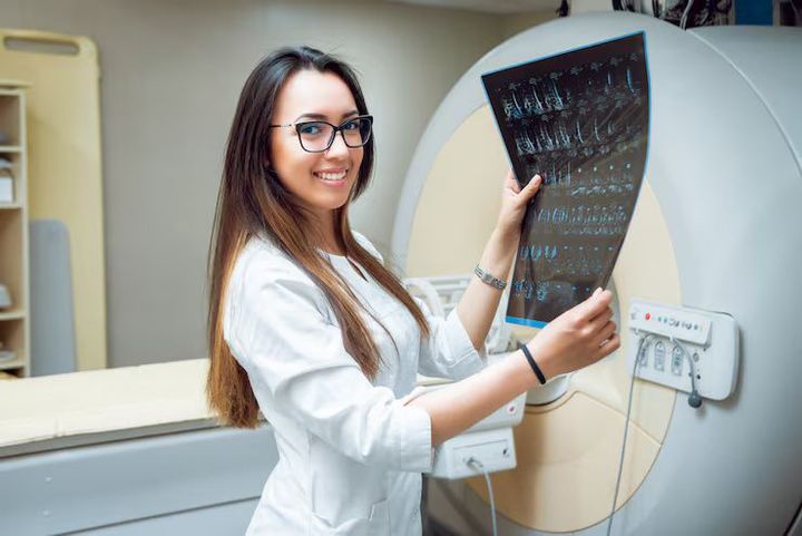
Medical imaging plays a critical role in modern healthcare by helping healthcare professionals evaluate injuries, diagnose medical conditions, monitor treatment progress, and support clinical decision-making.
Digital Radiography X Ray Machinery has transformed traditional X-ray imaging by replacing film-based systems with advanced digital detectors, allowing faster image acquisition, improved image quality, and more efficient workflow management.
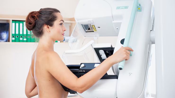
Hospitals, diagnostic imaging centers, clinics, dental practices, and emergency departments increasingly use digital radiography systems because they provide immediate image availability, support electronic record integration, and reduce the need for physical film processing. Understanding how digital radiography works can help healthcare organizations evaluate this important imaging technology.
Medical Disclaimer: This article is intended for general educational purposes only and should not replace professional medical advice. Medical imaging procedures should always be performed by qualified healthcare professionals using appropriate clinical guidelines and radiation safety practices.
What Is Digital Radiography X Ray Machinery?
Digital Radiography X Ray Machinery refers to X-ray imaging systems that capture images using digital detectors instead of traditional photographic film. The digital images can be viewed immediately, enhanced for diagnostic purposes, securely stored, and shared electronically with authorized healthcare providers.
Digital radiography is commonly used for:
Chest imaging
Bone and joint examinations
Orthopedic assessments
Emergency medicine
Dental imaging
Pediatric imaging
Routine diagnostic studies
Follow-up medical evaluations
The specific examination depends on the patient's medical needs.
How Digital Radiography Works
Digital radiography combines X-ray generation with advanced electronic image capture.
X-Ray Generation
The X-ray tube produces controlled X-ray beams that pass through the patient's body.
Image Detection
Digital detectors capture the transmitted X-rays and convert them into electronic signals.
Image Processing
Computer software processes the data to create high-resolution digital images.
Clinical Review
Healthcare professionals review the images using specialized diagnostic workstations.
Digital Storage
Images are securely stored within Picture Archiving and Communication Systems (PACS) or electronic health record systems where applicable.
Benefits of Digital Radiography
Digital imaging offers several advantages over traditional film-based methods.
Potential benefits include:
Faster image acquisition
Immediate image availability
Improved image quality
Digital image enhancement
Simplified electronic storage
Faster clinical workflow
Easier image sharing among authorized providers
Reduced need for film processing chemicals
Clinical outcomes depend on appropriate equipment use and professional interpretation.
Common Clinical Applications
Digital radiography supports a wide range of healthcare services.
Examples include:
Orthopedic Imaging
Evaluation of fractures, joint conditions, and musculoskeletal injuries.
Chest Imaging
Assessment of the lungs, heart, and surrounding structures.
Dental Radiography
Imaging of teeth, jaws, and oral structures.
Emergency Medicine
Rapid imaging for trauma assessment and urgent medical care.
Pediatric Imaging
Diagnostic imaging adapted to children while following radiation safety principles.
Surgical Planning
Supporting healthcare providers before and after certain medical procedures.
Key Features to Consider
Healthcare organizations often evaluate several factors when selecting digital radiography equipment.
Important considerations include:
Detector technology
Image resolution
Workflow integration
PACS compatibility
Radiation dose management features
Mobile imaging capabilities
Software functionality
Maintenance and technical support
Equipment selection depends on clinical requirements and facility needs.
Digital Radiography X Ray Machinery Overview
| Feature | Primary Benefit |
|---|---|
| Digital Detector | Immediate image capture |
| Image Processing Software | Enhanced diagnostic visualization |
| PACS Integration | Secure electronic image storage |
| High Resolution | Improved anatomical detail |
| Workflow Automation | Faster examination process |
| Dose Management Tools | Support optimized radiation practices |
Radiation Safety Considerations
Medical imaging professionals follow established radiation safety principles.
Helpful practices include:
Use imaging only when clinically appropriate.
Apply the lowest radiation dose necessary to achieve diagnostic image quality (often guided by the ALARA principle).
Shield patients when recommended and appropriate.
Maintain regular equipment calibration.
Train imaging personnel thoroughly.
Monitor equipment performance.
Follow applicable regulatory standards.
Document imaging procedures accurately.
Radiation safety is an important component of quality healthcare.
Current Industry Trends
Medical imaging technology continues to advance rapidly.
Recent developments include:
Artificial intelligence-assisted image analysis
Cloud-based image management
Wireless digital detectors
Mobile radiography systems
Automated workflow optimization
Enhanced cybersecurity for medical imaging
Predictive equipment maintenance
Integrated enterprise imaging platforms
These innovations improve workflow efficiency and support diagnostic decision-making.
Looking Ahead
The future of Digital Radiography X Ray Machinery is expected to include more advanced artificial intelligence tools, improved detector sensitivity, lower radiation dose technologies, expanded cloud connectivity, and deeper integration with electronic health systems. Continued innovation will further enhance imaging quality, operational efficiency, and patient care.
Why Understanding Digital Radiography X Ray Machinery Matters
Understanding Digital Radiography X Ray Machinery helps healthcare organizations evaluate imaging technologies that support accurate diagnosis, efficient clinical workflows, and secure digital image management. Proper equipment selection, routine maintenance, and adherence to radiation safety standards contribute to high-quality patient care.
Conclusion
Digital Radiography X Ray Machinery has revolutionized medical imaging by providing rapid, high-quality digital images that support timely diagnosis and effective clinical decision-making. Through advanced detectors, image processing software, electronic storage systems, and modern workflow automation, digital radiography improves efficiency across many healthcare settings. As imaging technology continues to evolve, digital radiography will remain a cornerstone of modern diagnostic medicine.